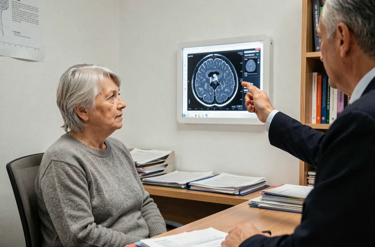
You receive an MRI report, scan the technical terms, and one phrase catches your eye: “Fazekas 1.” No context, no details about where the lesions are located, sometimes not even a comment from the radiologist beyond the score. This is a common situation, and the first reaction is often to search online for what the score means.
Grade 1 on the Fazekas scale corresponds to minor abnormalities in the brain’s white matter, visible as hyperintensities on MRI. In the vast majority of cases, this is a common marker of brain aging, not a disease in itself.
Fazekas 1 without further details: what the report may not tell you
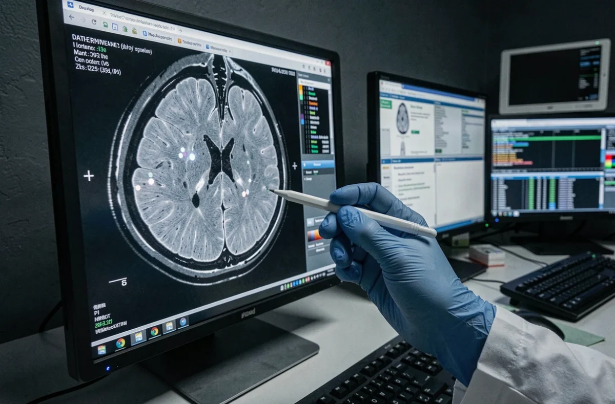
The Fazekas scale does not produce a single score. It distinguishes between two areas: the periventricular regions (around the brain’s central cavities) and the deep white matter. A periventricular grade 1 describes fine “caps” or rims along the ventricles. A deep grade 1 indicates a few small, scattered spots in the white matter.
The practical problem arises when the report mentions “Fazekas 1” without specifying which region is involved or which scoring convention was used. Some radiologists add the two subscores together, while others report the higher score of the two regions. The same “Fazekas 1” can therefore refer to different anatomical findings.
When faced with this lack of detail, a useful next step is to ask the referring doctor to review the report alongside the images, or to request clarification from the radiologist. A detailed article on the Fazekas scale and symptoms of Fazekas 1 can help you prepare these questions. Without clarification, you risk either downplaying a finding that warrants follow-up or worrying about an entirely unremarkable image.

White matter hyperintensities on MRI: a marker, not a diagnosis
One point that reports rarely make clear: a hyperintensity is a radiological finding, not proof of a specific cause. Recent research emphasizes the heterogeneous nature of these images. Their clinical significance depends on several factors considered together.
- The exact location of the lesions (periventricular, deep, or both) points to different mechanisms.
- Other abnormalities visible on the same MRI (microbleeds, lacunes, atrophy) affect how the grade is interpreted.
- The patient’s vascular risk profile (high blood pressure, diabetes, smoking) and any neurological symptoms determine the score’s real significance.
The Fazekas score describes the appearance and extent of the lesions. On its own, it is not enough to establish an underlying cause or predict an individual’s disease course. Fazekas 1 in a 70-year-old with high blood pressure does not have the same significance as in someone of the same age with no identified risk factors.
Variability between two MRI readings
Visual scoring involves some subjectivity. Studies report substantial agreement between readers, but not perfect agreement. Results also vary depending on the quality of the MRI sequences and the machine’s technical settings.
A small difference in grade between two consecutive scans does not necessarily mean there has been real progression. Volumetric methods (automated measurement of lesion volume) are more sensitive to subtle changes, but they are not interchangeable with the conventional visual score. In practice, it is better to compare the images side by side with the same radiologist than to rely solely on the number in the report.
Managing Fazekas 1: address vascular risk factors
There is no treatment that can make white matter lesions already visible on MRI disappear. Management therefore focuses not on the score itself, but on the factors that contribute to the development and progression of these lesions.
A 2023 consensus published in the Medical Journal of Australia on incidentally discovered hyperintensities recommends assessing and treating vascular risk factors according to standard guidelines. Controlling high blood pressure remains the main priority. Clinical trial data suggest that effective blood pressure reduction may slow the progression of hyperintensities in some patient groups.
The same consensus advises against starting an antiplatelet drug or anticoagulant solely because of these hyperintensities, unless there is another medical indication. In other words, an isolated Fazekas 1 finding does not, by itself, justify adding a medication.
Practical steps that make a difference day to day
Alongside medical follow-up, the steps you can take are the same as those used in standard vascular prevention, applied consistently.
- Monitor your blood pressure at home and keep it within the targets set by your doctor, adjusting treatment if needed.
- Stay physically active on a regular basis (brisk walking, swimming, cycling), which improves blood flow to the brain and reduces arterial stiffness.
- Manage diabetes or dyslipidemia according to current guidelines, without waiting for the lesions to progress.
- Quit smoking, a direct aggravating factor in cerebral small vessel disease.
It is worth asking your doctor about personalized blood pressure targets: the cognitive benefits of a lower target vary from patient to patient and cannot be determined from the MRI grade alone.
Fazekas 1 and symptoms: should you worry about cognitive decline?
Most people with Fazekas 1 have no neurological symptoms. Cognitive problems, difficulty walking, or changes in mood are more common at higher grades (Fazekas 2 and 3), when the lesions become confluent.
When symptoms occur in someone with a Fazekas 1 score, they often have another explanation: anxiety related to the diagnosis, sleep problems, medication side effects, or a separate associated condition unrelated to vascular white matter disease. Attributing cognitive problems to Fazekas 1 alone without further assessment would be a misinterpretation.
A follow-up MRI, scheduled one to three years later depending on the clinical context, can help confirm that the lesions are stable. If the grade remains stable and no symptoms develop, the outlook is favorable. In this scenario, Fazekas 1 remains what it most often is: a mark of time on the brain, like gray hair, warranting vascular vigilance but not alarm.